

You Need To Have A Yearly Exam When:
Your Medications Can Affect Your Eyes
You Have Diabetes, High Blood Pressure or Autoimmune Diseases
Family History Of Macular Degeneration or Glaucoma
Your Rx Is A High Myoptic Above (-4.00)
Medical Insurance Will Cover The Health Check Of The Eye.
Routine Health Checks For Eyes Begin Between 6-12 months And Again Before Starting School.
Ophthalmologist And Optometrist Need To Check For These Issues
If Your Doctor Is Not Checking-You Need To Ask
-

Eye Pressure
Intraocular pressure (IOP) is the fluid pressure inside the eye, primarily measured in the aqueous humor chamber. A healthy range is typically 10 mmHg to 20 mmHg. This pressure is regulated by a balanced flow: new aqueous humor is produced, and an equal amount drains out through the drainage angle. High IOP, called ocular hypertension, often results from the inadequate drainage of this fluid. Untreated high pressure severely stresses the eye's internal structures, particularly the optic nerve, which can lead to permanent damage, causing glaucoma and potential vision loss.
IOP is measured during an eye exam using tonometry, most commonly the non-contact "air puff" method, which gauges the force needed to flatten the cornea. If high IOP is detected, treatment is necessary to protect the optic nerve. This typically involves using prescription eye drops to either increase fluid drainage or reduce fluid production. In some cases, laser procedures or surgery may be required to maintain a safe pressure level and prevent the progression to glaucoma.
-
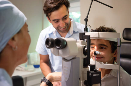
Slit Lamp-Biomicroscope
A slit lamp is a microscope with a bright light used during an eye exam. It gives your ophthalmologist a closer look at the different structures at the front of the eye and inside the eye. It’s a key tool in determining the health of your eyes and detecting eye disease.
Before the exam. There is no special preparation needed before a slit lamp exam.
The exam. Your doctor will have you sit in the chair in front of the slit lamp. You will be asked to place your chin in the chin rest and your forehead against the forehead band. This keeps your head steady during the exam. Your doctor may use eye drops that contain a yellow dye to help see problems with the front of the eye. Dilating drops may also be used to widen your pupil for a better look at the back of the eye. Your eye doctor will sit facing you and look through the microscope at your eyes. He or she will then turn on the slit lamp and focus a narrow, high-intensity beam of light towards your eye. Although the light is very bright, it will not cause any damage to your eye and should not cause any pain.
-
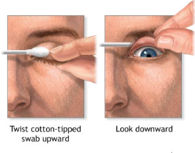
Lid Flip
Eyelid disorders that cause swelling, redness, or infection include:
Stye (hordeolum). A small, red, painful bump caused by a bacterial infection in an oil gland. A chalazion is a painless lump that may form after a stye.
Blepharitis. Makes your eyelids red, itchy, or burning and can cause crusty eyelashes. It's often linked to bacteria or skin conditions like rosacea.
Periorbital cellulitis. A bacterial infection of the eyelid and nearby skin. If it spreads behind the eye (orbital cellulitis), it needs immediate medical care.
Eyelid disorders that affect eyelid position or movement include:
Ectropion (eyelids turn out). The inner eyelid is exposed, which can cause irritation or tearing.
Entropion (eyelids turn in). The eyelashes rub against the eye, causing discomfort or damage.
Ptosis (drooping eyelid). Often caused by weak muscles, aging, or nerve damage.
Eyelid growths include:
Xanthelasma. Yellowish patches of cholesterol near the inner corners of the eyelids.
Papilloma. A benign (noncancerous), wart-like skin growth.
Basal cell carcinoma. The most common eyelid cancer, often on the lower eyelid.
Squamous cell carcinoma. A less common but faster-growing skin cancer.
Sebaceous gland carcinoma. A rare but serious cancer of the eyelid's oil glands.
Eyelid disorders that affect blinking or eyelid movement include:
Blepharospasm. Eyelid blinking or twitching you can't control.
Myokymia. A minor, occasional eyelid twitch often caused by fatigue or stress.